Gallbladder is an important organ of the digestive system. It is located posterior to the liver, about 3-4 inches long and acts as a storage unit of bile. Bile is a yellowish-green fluid produced by the liver that is vital for the digestion of fats or lipids. The gallbladder concentrates and holds the bile, till it is needed for fat digestion, upon receiving the signal for fat metabolism, it releases the bile into the duodenum.
The wall of the gallbladder has three layers, the innermost mucosal layer, the middle layer or the muscularis and the outer serosa.
Primary gallbladder cancer originates in the inner layer and spreads into the outer layers as it grows. It may also spread beyond that to affect the liver, bile duct, and stomach. 85% of Gallbladder Cancers (GBC) are adenocarcinoma; the remaining 15% are rare types of Gallbladder Cancers (GBC). There are three types of adenocarcinoma – Non-papillary adenocaricoma, papillary adenocarcinoma and mucinous adenocarcinoma. About 75% of all adenocarcinomas are non-papillary adenocarcinomas.
The other types of Gallbladder Cancers include – Squamous cell cancers, Adenosquamous Gallbladder Cancer, small cells Gallbladder Cancer, Sarcoma of the gall bladder, and NET (Neuroendocrinal tumor).
Facts about gallbladder cancer:
- It is a relatively rare cancer among GI (gastrointestinal) cancers. Yet, it constitutes about 80-90% of the cancers that affect the biliary tracts
- It is a malignancy with a marked ethnic and geographical variation (7/100,000 cases in India) versus the western countries (1.5/100,000 in the USA). Within India, it is more prevalent in the northern and the northeastern states (Bihar, Orissa, West Bengal)
- Women are twice more likely to be affected by it than men and the frequency increases in women above 65 years of age. Learn more.
- GBC (gallbladder cancer) is the most aggressive of biliary cancer, with a poor prognosis the survival rate of patients with gallbladder cancer in the advanced stage is a dismal 6 months. The 5-year survival rate is less than 5% in stage IV cancer.
- Gallbladder cancer incidence is low in south India and its association with gallstones is also low.
- Currently, there are no biochemical tests to detect GBC
What does gallbladder cancer feel like?
One of the challenges for early detection of gallbladder cancer is that gallbladder cancer symptoms can be vague and may mimic other conditions such as cholelithiasis and cholecystitis.
There are cases where gallbladder cancer has been incidentally diagnosed during a laparotomy – a surgical incision into the abdominal cavity in preparation for major surgery. By the time it presents in a late stage, the disease is characterized by local invasion, extensive regional lymph node metastasis, and distant metastasis or spread to other organs.
Gallbladder cancer has a high propensity to implant or seed onto peritoneum, laparoscopic or laparotomy wounds.
Since the gallbladder is located deep inside the body, cancer remains mostly undetected till it reaches its later stages. The scope of gall bladder treatment increases if detected early.
Signs and symptoms of gallbladder cancer:
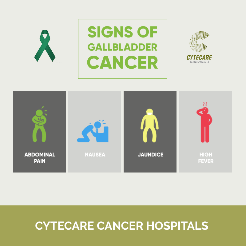
- Pain in the upper right part of the belly
- Gall bladder enlargement
- Jaundice, with yellowing of the skin and in the white part of eyes (caused as a result of bile accumulation in different body parts)
- Swelling in and around the belly
- Other symptoms include nausea, loss of appetite, weight loss, fever, itchiness in the skin, etc.
Advanced stage gallbladder cancer symptoms:
- Jaundice – yellowing of the skin and whites of the eyes
- Steady abdominal pain, especially in the upper right region
- Nausea, bile, and vomiting
- Indigestion
- Bloating
- A lump in the abdomen
- Fever
- Loss of appetite
- Weakness
- Unnatural weight loss
Can gallbladder cancer be seen on ultrasound?
An ultrasound would help to detect any tumours present in the gallbladder walls, or its spread if that caused the lymph nodes to enlarge. An endoscopic ultrasound might help to detect and differentiate between a benign vs malignant polyp.
The detection of gallbladder stones if entrapped in a mass becomes challenging to diagnose with ultrasound, and there are chances to miss the cancer growth. Other diagnostic tests will help in GBC detection.
Diagnostic tests for gallbladder cancer:
Currently, there are no biochemical tests for the early detection of gallbladder cancer. One of the first modalities for detection is a transabdominal ultrasound (USG). However, USG is user-dependent and subtle changes in the gall bladder could be missed.
Imaging tests provide better resolution and are helpful in identifying structural changes to the gallbladder induced by the tumor, such as mass, polyps, parietal thickening, etc. Imaging modalities such as CT, PET, MRI, ERCP (endoscopic retrograde cholangiopancreatography) MRCP (magnetic resonance Cholangiopancreatography) are used for detection as well as the staging of the tumor.
Often laparoscopic biopsy is obtained instead of FNAC (fine needle aspiration) in case of gallbladder cancer as it has a high propensity for seeding. Information on the stage and the pathology of the tumor will be critical in addressing the next steps of treatment in gallbladder cancer.
It is important to remember that people with gallstones need to be monitored closely for gallbladder cancer.
Gallbladder cancer risk factors:
The following factors can raise a person’s risk of developing this cancer:
- Age – It usually occurs in older people, over the age of 45 years
- Gender – The cancer has shown strong links to the female gender
- Ethnicity – Certain ethnic groups have been observed to be more susceptible to gallbladder cancer. These include Mexican Americans, Native Americans, Indians, Pakistan, people in certain regions of East Europe, East Asia and South America.
- Gallstones – This is considered to be the most common risk factor of gallbladder cancer
- Gallbladder polyp – A type of growth that sometimes forms when small gallstones get embedded in the gallbladder wall
- Poor lifestyle– Smoking, being overweight, imbalanced diet, and suffering from chronic infections of gallbladder
What is the survival rate of gallbladder cancer?
The 5-year survival rate of gallbladder cancer is less than 5% in the country.
This low survival rate comes with the fact that for most patients, this cancer remains undetected until it reaches its last stage, which reduces the surgical treatment of the tumour that has spread.
Is gallbladder cancer curable?
Gallbladder cancer is curable when detected in its early stages.
When in its later stages, the patient gets palliative and support care, but with the difficulty of its early detection, gallbladder cancer remains incurable without its surgical removal.
Gallbladder cancer treatment options:
Gallbladder cancer has a poor outcome and prognosis. When detected in an early stage, complete surgical resection of the gall bladder is the best option. Surgical management of gallbladder cancer is dependent on the extent of the spread of gallbladder cancer (to liver, intra-peritoneal spread, lymphatic and vascular invasion).
Depending on the stage of the tumor, the primary gallbladder cancer treatment may involve simple cholecystectomy or radical surgery, involving removal of the gall bladder, liver resection (hepatectomy) or removal of sections of the liver, and lymph nodes. If there is a pancreatic or duodenal involvement, additional resection involving the pancreas and the duodenum may be performed (Hepato-pancreaticoduodenectomy, excision of duodenum wall in localized involvement, Pancreaticoduodenectomy for local and peri pancreatic lymph spread).
Radiation therapy modalities including external beam therapy, intraoperative radiotherapy, and brachytherapy with or without chemotherapy have been used for adjuvant treatment of gallbladder cancer. Adjuvant treatment is often used in all patients diagnosed post stage II to IVA.
The benefit of adjuvant chemotherapy in gallbladder cancer has been observed in node-positive patients. Some of the common chemotherapeutic agents used for the treatment of gallbladder cancer include-Gemcitabine, Cisplatin, 5-fluorouracil, cepacitabine.
In the case of gallbladder cancer, Chemotherapy is directly injected into the hepatic artery (hepatic artery infusion) unlike injecting it into a vein, to maximize the benefit of chemotherapy. Since the hepatic artery also supplies to the gall bladder tumor, the drug can be effectively delivered in concentration to the tumor. Chemotherapy may also be useful in the management of unresectable tumors.
Preventive measures for gallbladder cancer
While there is no sure way of gallbladder cancer prevention, and one cannot control natural factors like age, gender, and ethnicity, it is recommended that one lead an active and healthy lifestyle. A healthy diet with cereals, whole grains instead of refined, at least two and a half cups of fruits and vegetables daily, and limiting the intake of processed food and red meat have been shown to lower the risk of many cancers.


